Introduction
Salvage radiotherapy (RT) to the prostatic fossa is an effective therapy in patients with biochemical or local recurrence after radical prostatectomy (RP). The American Society for Radiation Oncology (ASTRO) and the American Urological Association (AUA) guidelines recommended salvage RT for treatment of biochemical or local recurrence without evidence of distant metastasis [1]. Several randomized trials have included a subgroup with detectable prostate-specific antigen (PSA) levels after RP. RT after RP significantly reduced local failure and distant metastasis and improved overall survival in the Southwest Oncology Group (SWOG) 8794 [2,3] and biochemical failure in the European Organisation for Research and Treatment of Cancer (EORTC) 22911 [4] among patients with postoperative high-risk factors or a detectable PSA level. Two observational studies have reported the outcomes of salvage RT. At a median of 11.5 years, salvage RT after RP significantly reduced the local recurrence risk by almost 90% and systemic progression by 75% [5] and was associated with a three-fold increase in prostate cancer-specific survival relative to those who received no salvage treatment [6].
Although salvage RT is the only potentially curative treatment option for recurrent prostate cancer patients after RP, the optimal target volume of salvage RT remains controversial. Several guidelines have been published for clinical target volume (CTV) delineation in postoperative RT [7-10]; however, there is no universally accepted method, and substantial variation exists.
In the postoperative setting, anatomic changes that occur following RP may significantly alter the target definition compared with definitive RT for localized prostate cancer patients. The absence of a visible target introduces a potentially significant interobserver bias in defining the prostatic fossa CTV.
Magnetic resonance imaging (MRI) provides greater resolution of soft tissues compared with computed tomography (CT), and it allows more precise delineation of target volume and normal tissues [11,12]. Therefore, MRI is commonly used in the planning of definitive RT for prostate cancer. Several studies have reported that addition of MRI to planning CT decreases target volume and reduces variation in prostate delineation [13,14]. Also, MRI has the potential to demonstrate the site of local recurrence after RP [7-9] and can be used for CTV delineation in salvage RT.
In this study, we investigated interobserver variation in target volume delineation for salvage RT in recurrent prostate cancer patients treated with radical prostatectomy, and we compared the effect of addition of MRI in determination of CTV.
Materials and Methods
1. Case and observer characteristics
Planning CT scans of five sampled cases referred for salvage RT to the prostatic fossa because of continuous PSA elevation after RP were used for this target delineation study. Each case involved diagnostic prostate MRI before RP and planning MRI before salvage RT. The clinical and surgical information of the cases is shown in Table 1.
Ten radiation oncologists from 10 different institutions participated in the study and were asked to contour the target volume on each of the five CT scans. Their institutions’ practice patterns for salvage RT are described in Table 2.
2. Planning CT and MRI acquisition
The planning CT and MRI scans were performed in a reproducible supine position with endorectal ballooning at same day. All patients voided their bladders immediately before scanning. Treatment planning CT scans of the pelvis were obtained in 2.5-mm slice thicknesses at 2.5-mm intervals using a LightSpeed RT16 scanner (GE Healthcare, St. Giles, UK). The MRI scans were performed with 3.0T systems (Achieva 3T, Philips Medical System, Best, Netherlands) using a 6-channel phased-array coil. Both T2-weighted images (T2WIs) and diffusion-weighted images (DWIs) were included in all of the MRI scans. The T2WIs were acquired in the three orthogonal planes (axial, sagittal, and coronal). The following T2WI scan parameters were used for this study: repetition time (TR)/echo time (TE), 2,600-4,200 ms/80–100 ms; slice thickness, 3 mm; interslice gap, 0.3–0.1 mm; matrix, 512×304; field-of-view (FOV), 15–20 cm; number of signals acquired, 3; and sensitivity encoding (SENSE) factor, 2. The DWIs were acquired in the axial plane using the single-shot echo-planar imaging technique. The scanning parameters were as follows: TR/TE, 2,740–2,750 ms/83–85 ms; slice thickness, 3 mm; interslice gap, 1 mm; matrix, 112 × 110; FOV, 20 cm; SENSE factor, 2; and the number of signals averaged (NSA), 3.
3. Target volume delineation
Ten observers received the clinical information and pathological status of five cases in order to define CTV (Table 1). The planning CT and diagnostic MRI (T2WI and DWI) scans were offered to each observer using an online storage system and were transferred to each individual planning system. On every CT slice, the first contouring of CTV was performed under institutional practice guideline whether or not diagnostic MRI was performed. After an interval of 2 weeks, the planning MRI scans (T2WI with endorectal ballooning and DWI) of each case were offered and superimposed on the CT image. The second contouring of CTV was drawn on superimposed CT and axial MRI slices of each scan. All CTVs included only the prostate fossa (pelvic lymph node area was excluded).
4. Analysis of interobserver variation
After completion of contouring, planning CT images with two different CTVs were analyzed using the Pinnacle3 planning system 9.2 (Philips Medical Systems, Fitchburg, WI, USA). Data on each delineated target volume both on planning CT only (CTV1) and on CT with planning MRI (CTV2) were imported from the RT planning system. For each case, the average volume and standard deviation (SD) of the ten observer measurements were calculated. Subsequently, the mean of these averages and their SDs were calculated for both CTV1 and CTV2. Comparisons between CTV1 and CTV2 derived values (mean or SD) were represented in percentage decrease, defined as (xCTV2–xCTV1)/xCTV1; positive values indicated that the CTV2 value was larger than the corresponding CTV1 value.
Statistical analysis was executed using SAS ver. 9.3 (SAS Institute, Cary, NC, USA). The Wilcoxon signed-rank test was used to compare measurements of target volume. A linear mixed model was used to identify the factors that influenced target volume delineation. All p-values were two-sided, and those less than 0.05 were regarded as statistically significant.
Results
1. Interobserver variations in CTV1 and CTV2
There were large differences in CTV delineated by each observer, regardless of the addition of planning MRI. Table 3 shows the CTVs determined by CTV1 and CTV2 for each case. Between the observers, 7- to 15-fold differences existed.
2. Target volume change using planning MRI
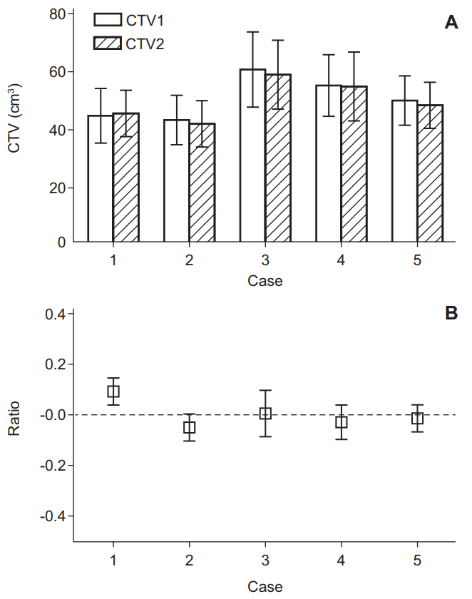
There were no significant differences in mean and SD between CTV1 and CTV2 (50.6 ± 31.6 cm3 in CTV1 and 49.7 ± 30.6 cm3 in CTV2; p = 0.1875). The mean CTV2 of the observers decreased compared with CTV1 for all except case #1. The SD of CTV2 was also decreased compared with CTV1 for all except case #4 (Fig. 1A). The percentage decrease in the CTVs is demonstrated in Fig. 1B. The observers tended to report a decreased mean volume and SD as a result of using the planning MRI.
3. CTV1 and CTV2 difference according to observers
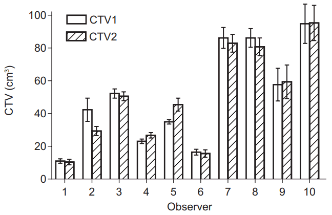
The target volumes were also analyzed by each observer. Fig. 2 shows mean CTVs and standard errors of each observer. Observer 10 reported the largest volume (94.7 ± 27.2 cm3 in CTV1 and 95.1 ± 24.6 cm3 in CTV2), and observer 1 reported the smallest volume (11.3 ± 1.5 cm3 in CTV1 and 10.6 ± 2.4 cm3 in CTV2). Six observers delineated the decreased CTV (CTV2) using planning MRI.
Discussion and Conclusion
In adjuvant or salvage RT after RP, there is no optimal consensus for CTV delineation. Several guidelines have been published based on the pattern of local failure. Malone et al. [15] compared four of the guidelines [7-10] and reported that the recommended CTV was significantly different among the guidelines. The CTVs proposed by the EORTC, the Faculty of Radiation Oncology Genito-Urinary Group (FROGG), the Radiation Therapy Oncology Group (RTOG), and the Princess Margaret Hospital (PMH) differ based on the recommended target border, especially in the cranial direction. The superior boundary of the CTV was defined as follows: by the bladder neck in the EORTC guideline; as encompassing the entire seminal vesicle bed and distal portion of the vas deferens in the FROGG guideline; as the level of the cut end of the vas deferens or 3–4 cm above the top of the symphysis in the RTOG guideline; and as the superior surgical clips or 5 mm above the vas deferens in the PMH guideline. The volumes of the four guidelines were 60 ± 17 cm3, 88 ± 16 cm3, 102 ± 24 cm3, and 104 ± 25 cm3, respectively.
Recently, we analyzed the recurrent tumor location in 113 prostate cancer patients using MRI and suggested an optimal CTV, which included 97% of suspected tumor recurrences. Using the inferior border of the pubic symphysis as a point of reference, the inferior border of the CTV was 8 mm below and the superior border extended 30 mm above. We suggested that it might be unnecessary to routinely include the seminal vesicle bed or the surgical clips within the CTV for all patients. With this guideline, the mean CTV in our practice was 15 ± 5 cm3, remarkably smaller than those of the other guidelines [16].
In this study, there were large differences in CTV, regardless of planning MRI fusion between observers. These differences were due to the differing CTV delineation guidelines among the different institutions. Our results indicate that CTV delineation by fusion of planning CT and planning MRI might reduce clinical target volume.
The organ-discriminating power on CT images is much lower than that on MR images. CT can be used to discriminate various tissues based solely on differences in attenuation coefficients [10]. Since the prostate, rectal and bladder wall, pelvic floor muscles, and penile bulb have similar attenuation coefficients, they cannot readily be discriminated on CT images, resulting in potentially significant inaccuracies. Conversely, MRI can be used to demonstrate and characterize soft tissues by providing superb soft-tissue contrast on T2WIs. MRI therefore shows much more detail on the prostatic margins in any direction, leading to more accurate delineations. Compared with MRI, CT images overestimate prostate volume by 35% [14]. Villeirs et al. [13] reported that using CT plus MRI compared to CT alone resulted in significant decreases of 6.54%, 5.21%, and 10.47% in mean CTV of prostate and seminal vesicle volumes, and the SDs were significantly decreased by 63.06%, 62.65%, and 44.83%, respectively. These results indicate that the addition of MRI to CT decreases the interobserver target volume delineation variation in definitive RT for prostate cancer. Therefore, this greater resolution of soft tissues through the use of MRI could help the delineation of CTV for salvage RT through the anatomic precision of prostatic fossa.
Despite a substantial difference between CTVs delineated by each observer, a trend of volume reduction was observed in the mean CTV and SD when planning MRI was added. When we considered salvage RT procedures of 10 institutions, the observers using endorectal ballooning and bladder filling in clinical practice affected the CTV and had a tendency of decreasing target volume with MRI. Anatomic accuracy of prostatic fossa using planning MRI might be one explanation for the results.
In conclusion, we show that interobserver variation in target volume delineation for salvage RT is substantial. The combination of planning MRI with CT tends to decrease the target volume and its variation. However, the role of MRI in salvage RT target delineation was insufficient, and additional studies are needed. Furthermore, to reduce interobserver variation, a consensus meeting for optimal target volume after RP is needed among physicians.