Treatment results of radiotherapy following CHOP or R-CHOP in limited-stage head-and-neck diffuse large B-cell lymphoma: a single institutional experience
Article information
Abstract
Purpose
This study evaluated outcomes of radiotherapy (RT) after chemotherapy in limited-stage head-and-neck diffuse large B-cell lymphoma (DLBCL).
Materials and Methods
Eighty patients who were treated for limited-stage head-and-neck DLBCL with CHOP (n = 43) or R-CHOP (n = 37), were analyzed. After chemotherapy, RT was administered to the extended field (n = 60) or the involved field (n = 16), or the involved site (n = 4). The median dose of RT ranged from 36 Gy in case of those with a complete response, to 45–60 Gy in those with a partial response.
Results
In all patients, the 5-year overall survival (OS) and disease-free survival (DFS) rates were 83.9% and 80.1%, respectively. In comparison with the CHOP regimen, the R-CHOP regimen showed a better 5-year DFS (86.5% vs. 73.9%, p = 0.027) and a lower rate of treatment failures (25.6% vs. 8.1%, p = 0.040). The volume (p = 0.047) and dose of RT (p < 0.001) were significantly reduced in patients treated with R-CHOP compared to that in those treated with CHOP.
Conclusion
The outcomes of RT after chemotherapy with R-CHOP were better than those of CHOP regimen for limited-stage head-and-neck DLBCL. In patients treated with R-CHOP, a reduced RT dose and volume might be feasible without increasing treatment failures.
Introduction
In Korea, 4,553 patients were treated for non-Hodgkin’s lymphoma (NHL) in 2012; this condition ranked as the malignant tumor with the 10th highest incidence rate of all cancers. The incidence rate of NHL was reported to be 6.6% in 2012, but is increasing in Korea [1]. Among all NHL cases, the head and neck region was involved in about 15% of cases [2]. Radiotherapy (RT) has been widely used after chemotherapy in the treatment of NHL, and some studies showed that reducing the volume and dose of RT doesn’t affect the treatment results [2-5]. According to the International Lymphoma Radiation Oncology Group (ILROG) recommendation, the volume could be limited to the involved lesion and the dose could be reduced to 30 Gy by response after chemotherapy in patients with head and neck lymphoma [6]. The addition of R-CHOP (rituximab to cyclophosphamide, doxorubicin, vincristine, and prednisone) could improve clinical outcomes [7-9], but it is unclear that it could be better outcomes in patients treated with RT after R-CHOP.
The purpose of this study is to identify whether the volume or dose of RT and the regimen of chemotherapy affect treatment outcomes in patients with diffuse large B-cell lymphoma (DLBCL) limited to the head and neck region. In present study, we included the limited-stage head-and-neck DLBCL patients treated with chemotherapy, CHOP or R-CHOP, followed by RT.
Materials and Methods
We identified 92 patients with limited-stage DLBCL of the head-and-neck region, who received RT at Chonnam National University Hwasun Hospital, from January 1994 to December 2010. Among these patients, we reviewed 80 patients who treated with RT after chemotherapy. The institutional ethical committee of the Chonnam National University Hwasun Hospital approved this study (No. CNUHH-2015-057).
The diagnoses of all patients were pathologically confirmed at local clinics or at our institution. Routine work-up studies of patients included a past history of systemic disease, B-symptoms, physical examination, complete blood counts and peripheral blood smear, serum lactate dehydrogenase (LDH) levels, and evaluation of renal and liver functions. Other staging procedures included bone marrow biopsy, chest radiographs, computed tomography (CT) of the neck, chest, abdomen, and pelvis. Some of the patients underwent positron-emission tomography/computed tomography (PET/ CT). Disease stage was determined based on the Ann Arbor Staging Classification [10].
All patients were treated with chemotherapy. From 1994 to 1999, three to six cycles of chemotherapy of CHOP (cyclophosphamide, vincristine, doxorubicin, and prednisolone) followed by extended field RT (EFRT) for the whole neck lymph nodes (LN) including Waldeyer’s ring and the supraclavicular region were recommended for limited stage DLBCL. Since 2000, chemotherapy of rituximab and CHOP (R-CHOP) followed by involved-field RT (IFRT) to ipsilateral involved LN and adjacent LN has been preferred. Some patients received involved-site RT (ISRT) to the pre-chemotherapy gross tumor with a 1 cm margin [6]. In patients treated with EFRT or IFRT, radiation field was confined to involved site after elective nodal irradiation of 30–45 Gy in 1.8 Gy or 2.0 Gy per daily fraction. The three-dimensional conformal RT or conventional RT using 6 MV photon beams were performed by linear accelerator.
All patients underwent an initial CT scan at diagnosis, and a subsequent interim CT after the third or fourth cycle of chemotherapy to evaluate the response to therapy. Several patients underwent PET/CT at diagnosis and after treatment. The final response was assessed within 1 month after completion of the chemotherapy and RT. A complete response (CR) was defined as complete disappearance of all clinical or radiological evidence of disease, and a partial response (PR) was defined as a tumor decreased to less than 50% on radiological imaging. In patients underwent PET/CT, CR was defined as negative fluorodeoxyglucose (FDG) uptake in a PET-positive tumor prior to R-CHOP regardless of residual lesion on CT according to consensus guideline by International Harmonization Project in Lymphoma [11]. In cases of variable FDG-avid or FDG-avidity uncertain tumors, CR was defined as disappearance of extranodal lesion or regression of nodal lesion to normal size on CT. Patients underwent a follow-up restaging examination every 3 months during the second year after treatment, and every 6 months thereafter. This included routine blood test, CT of the head-and-neck, chest, abdomen, and pelvis.
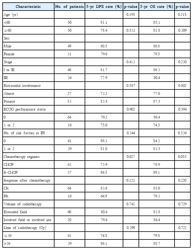
The following variables were included in the analysis of prognostic factors for overall survival (OS) and disease-free survival (DFS): age (<60 vs. ≥60 years), sex, stage (I vs. II), extranodal involvement (absence vs. presence), the Eastern Cooperative Oncology Group (ECOG) performance status (0 vs. 1 or 2), number of risk factors according to the International Prognostic Index (IPI) (0 vs. 1 or 2), chemotherapy regimen (CHOP vs. R-CHOP), response to chemotherapy (CR vs. PR), volume of radiotherapy (extended field vs. IFRT or ISRT), and dose of radiotherapy (<36 Gy vs. ≥36 Gy). The Kaplan-Meier method was used to plot survival curves. Univariate analysis by the log-rank test was performed to identify prognostic factors. To evaluate factors predictive of treatment failure, we used chi-squared tests and Student t-test.
We evaluated OS and DFS as end-points, using Kaplan-Meier methods. The duration of OS was measured from the date of the start of chemotherapy until the date of death from any cause or last follow-up. The duration of DFS was measured from the date of the start of chemotherapy until the date of the first recurrence of lymphoma or death due to any cause. Statistical analysis was performed using SPSS ver. 21.0 software (IBM, Armonk, NY, USA).
Results
Of 80 patients, 49 patients (61.3%) were male, with a median age of 54 years. Patients were grouped according to the Ann Arbor Staging system as follows: 46 (57.6%) patients, stage I; and 34(42.4%) patients, stage II. Most patients presented with ECOG 0 or 1 (98.7%), and without B-symptoms (96.3%).
Six of 80 patients (7.5%) had a bulky mass more than 6 cm in the longest diameter on CT imaging. Of 80 patients, 53 (66.2%) revealed various sites of extranodal presentation. The tonsil was the most common site of extranodal involvement and 42 patients had only tonsillar lesions. Other involved sites were as follows: the nasal cavity in 2 patients, the nasal cavity and nasopharynx in one, nasopharynx only in one, oropharynx in one, oropharynx and tonsils in one, oropharynx and tongue base in one, uvula in one, buccal mucosa in one, epiglottis in one, arytenoids in one, respectively. Chemotherapy was performed for three to six cycles of CHOP in 43 of 80 (53.8%) patients, and with R-CHOP in 37 patients, respectively (Table 1).
After completion of chemotherapy, 64 patients (80.0%) demonstrated a CR and 16 (20.0%) a PR. Of 43 patients who received CHOP, 31 patients (72.1%) showed CR. And among 37 patients who received R-CHOP, 33 patients (89.1%) showed CR. Radiation therapy was conducted with three-dimensional technique in 30 patients (37.5%) and conventional technique in 50 patients (62.5%). Median doses of RT were 36 Gy (range, 23.4 to 50 Gy) in patients with CR, and 45 Gy (range, 36 to 60 Gy) with PR after chemotherapy. Sixty patients (75.0%) received extended EFRT, 16 received IFRT, and 4 received ISRT. After completion of RT, all patients showed a CR.
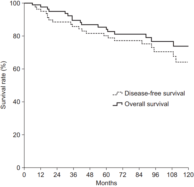
The median follow-up period was 85 months (range, 6 to 265 months). The 5-year OS and DFS of 80 patients were 83.9% and 80.1%, respectively (Fig. 1). The patients who received R-CHOP showed a significantly higher 5-year DFS (86.5%) compared to those who received CHOP (73.9%) (p = 0.027). The patients who received R-CHOP showed a higher 5-year OS (89.1%) compared to those who received CHOP (78.9%) with marginal significance (p = 0.055) (Table 2, Fig. 2).
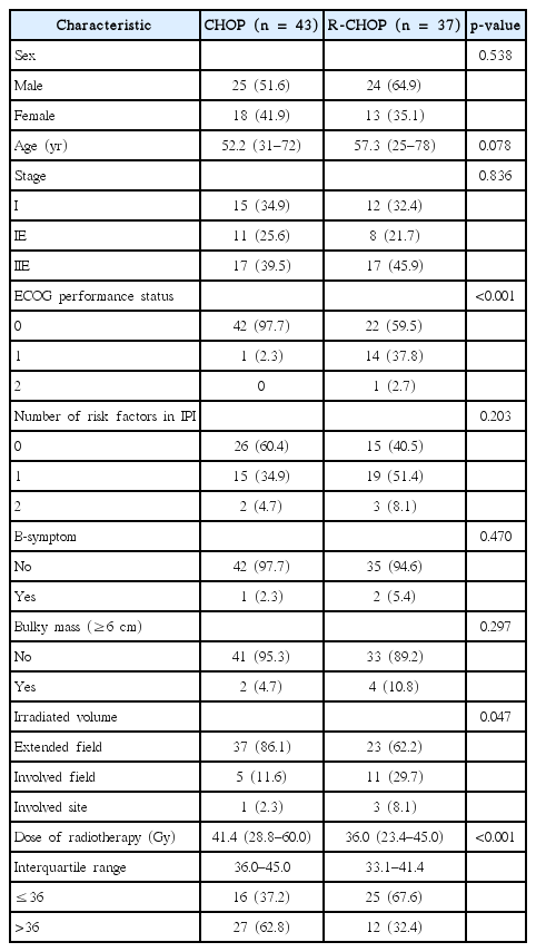
Fourteen patients (17.5%) showed treatment failure (Table 3). Two patients (2.5%) had isolated local recurrences, 11 (13.8%) had isolated distant recurrences, and 1 (1.2%) had simultaneous local and distant recurrence. The most common site of distant metastases was axillary LN (5 of 12 patients). Other sites of distant metastases were mediastinal LN (n = 1), abdominal LN (n = 2), inguinal LN (n = 1), eyelid (n = 1), breast (n = 1), chestwall (n = 1), liver (n = 1), spleen (n = 1), ascending colon (n = 1), duodenum (n = 1), ileum (n = 1), and bone (n = 1). All local recurrences were occurred in the field of radiation therapy (in-field failure). Ten of 60 patients (16.7%) who had received EFRT, and 4 of 20 patients (20.0%) who had received IFRT or ISRT, experienced treatment failure (p = 0.734). The dose of RT was lower in the patients with treatment failure (median, 36 Gy) than those without treatment failure (median, 39.6 Gy), but not significant (p = 0.094). There was no significant difference between the two groups of CR and PR after chemotherapy in treatment failures (p = 0.883). The chemotherapy regimen used was a significant predictive factor related to treatment failure (p = 0.040), 11 patients who had received CHOP (25.6%) and three who had received R-CHOP (8.1%) showed treatment failure.
The volume (p = 0.047) and dose of RT (p < 0.001) were significantly reduced in patients who had received R-CHOP compared to that in those who had received CHOP. Twenty-seven patients in CHOP (62.8%) and 12 in R-CHOP (32.4%) treated with RT more than 36 Gy, respectively (p = 0.007) (Table 4). However, the dose of RT was not a significant factor for OS (p = 0.909) or DFS (p = 0.952) in patients with CHOP. Also, it didn’t affect significantly on OS (p = 0.911) or DFS (p = 0.821) in patients with R-CHOP.
Discussion and Conclusion
Treatment of chemotherapy followed by RT has been used in patients with limited stage DLBCL. There have been several prospective studies of combined chemotherapy, CHOP, and RT that have demonstrated better outcomes than chemotherapy alone in patients with limited stage DLBCL. Miller et al. [12] have reported a better 5-year progression-free survival (PFS) (77% vs. 64%, p = 0.03) and OS (82% vs. 72%, p = 0.02) rate with three cycles of CHOP and RT than with eight cycles of CHOP alone in patients with stage I or II DLBCL. Moreover, in a randomized study comparing consolidation RT with observation after eight cycles of CHOP for patients with stage I or II DLBCL [13], the 6-year DFS was significantly different (73% for RT and 56% for observation; p = 0.05). Present study revealed similar results of CHOP followed by RT, in that the 5-year DFS and OS were 73.9% and 78.9%, respectively.
Addition of rituximab to CHOP improves the treatment outcomes in patients with DLBCL [7-9]. However, it remains questionable whether addition of rituximab to CHOP could improve treatment outcomes when RT is performed after chemotherapy because there were no randomized trials. Several retrospective studies reported that combination with RT and R-CHOP could improve the treatment outcome. In a study for patients with stage I-IV DLBCL treated with R-CHOP, significant improvements in 5-year OS (91% vs. 83%, p = 0.015) and PFS (90% vs. 75%, p < 0.001) were reported in patients who received RT compared to those who did not receive RT [14]. Among those patients with stage I or II DLBCL, patients who received RT had a longer OS (HR = 0.52) and PFS (HR = 0.45) than those who did not receive RT. Kwon et al. [15] reported that 3-year PFS and OS were 92.7% and 95.0% in stage I or II DLBCL patients treated with RT after R-CHOP. Additional RT was a significant prognostic factor in PFS (HR = 0.23; 95% CI, 0.07–0.80; p = 0.021) and OS (HR = 0.15; 95% CI, 0.03–0.68; p = 0.014) in multivariate analysis. In our study, patients who received R-CHOP showed similar results of R-CHOP followed by RT, in that the 5-year DFS and OS were 86.5% and 89.1%, respectively.
The optimal regimen of chemotherapy in patients with limited-stage DLBCL receiving RT is unclear. In a retrospective study of limited-stage head-and-neck DLBCL patients [2], the 5-year OS and PFS were 89.1% and 86.7% for patients receiving RT after CHOP and 100% each for those receiving R-CHOP, but there was no statistically significant difference. In our study, patients who received R-CHOP showed a significantly superior outcome in the 5-year DFS as compared to those receiving CHOP (86.5% vs. 73.9%, p = 0.027). Although there was no randomized trial, R-CHOP followed by RT is recommended in patients with limited-stage DLBCL [16] on the basis of which R-CHOP have been shown more effective than CHOP [7-9].
There is currently no consensus on the adequate RT volume that should be used for localized DLBCL. Traditionally, IFRT was used to treat Hodgkin’s lymphoma or NHL, which included the site of the clinically involved lymph node group [17]. Nathu et al. [18] reported treatment failures after RT in patients with stage I or II NHL of head-and-neck. They reported that 90% of failures outside of RT volume occurred in cervical lymphatic regions, such as Waldeyer’s ring and the supraclavicular region. Recently, there have been some reports to suggest effective local control could be achieved with a reduced volume of RT. Yu et al. [2] reported results of involved ISRT after chemotherapy by CHOP or R-CHOP in limited-stage head-and-neck DLBCL. The 5-year OS and PFS were 89.2% and 88.9%, respectively, with 8 treatment failures. Among these patients, only one patient experienced recurrence inside of the RT field. In a retrospective study for patients with limited-stage DLBCL [3], the time-to-progression (p = 0.823), PFS (p = 0.575), and OS (p = 0.417) were not significantly different between IFRT and ISRT delivered to the pre-chemotherapy-involved node with a margin ≤5 cm. Another study showed that IFRT resulted in a significantly higher incidence of higher grade toxicities than did ISRT, with no significant difference in treatment outcomes [19]. In our institution, 60 patients received EFRT, 16 received IFRT, and 4 received ISRT. The irradiated volume did not affect the DFS and OS significantly. Reduced-field irradiation could thus be feasible in patients with limited-stage DLBCL involving the head-and-neck.
There is no uniform dose of RT used in DLBCL. Historically, the RT dose ranged from 40 to 55 Gy to the gross lesion [12]. There has been a trend to reduce the RT dose when combined with chemotherapy. A randomized trial used 30 Gy in cases of CR and 40 Gy in cases with PR after CHOP in patients with limited-stage DLBCL [13]; the 6-year DFS and OS were 73% and 82%, respectively. In a retrospective study using R-CHOP and RT in DLBCL patients, the dose ranged from 30 to 39 Gy [14]. The 5-year OS and PFS were 91% and 82%, without relapse in the RT field. Krol et al. [4] reported no statistically significant differences in the OS and DFS between stage I DLBCL patients receiving 24 or 40 Gy who had reached a CR after CHOP. In a randomized trial comparing 45 to 30 Gy dose in patients with NHL, no statistically significant differences in the within-radiation field progression, PFS, and OS were observed [5].
Wilder et al. [20] investigated the relationship between pre-chemotherapy tumor size and the dose of radiation delivered in patients with intermediate-grade and large-cell immunoblastic lymphoma who demonstrated a CR after chemotherapy. They found that a tumor size less than 3.5 cm had excellent local control with low doses of 29.1-39.1 Gy, and tumors of 3.5-10 cm achieved better local control with higher doses of 39.2-50.8 Gy. In a guideline by the National Comprehensive Cancer Network (NCCN) or ILROG, the dose of 30-36 Gy was recommended for consolidation RT after chemotherapy, and a dose of 40-45 Gy was recommended for complementary RT for residual lesion [6,16]. In present study, the dose of RT was similar to these recommendations, and did not affect DFS and OS. After chemotherapy, a dose of 30–36 Gy could be sufficient to prevent treatment failures in patients with CR. And boost could be added to residual lesion after RT of 36 Gy.
This study has a several limitations. Its retrospective nature may create the risk of selection bias; and the relatively small number of cases may be insufficient to derive conclusions. Nevertheless, this study revealed that RT after R-CHOP results in a better prognosis than RT after CHOP, in spite of recued dose and volume of RT, in patients with limited-stage head-and-neck DLBCL. Use of a reduced volume and low dose of 36 Gy of RT could be feasible. In the future, a prospective randomized study is necessary.
Notes
Conflict of Interest
No potential conflict of interest relevant to this article was reported.


